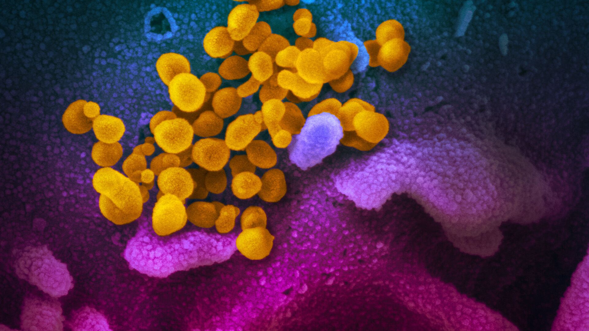
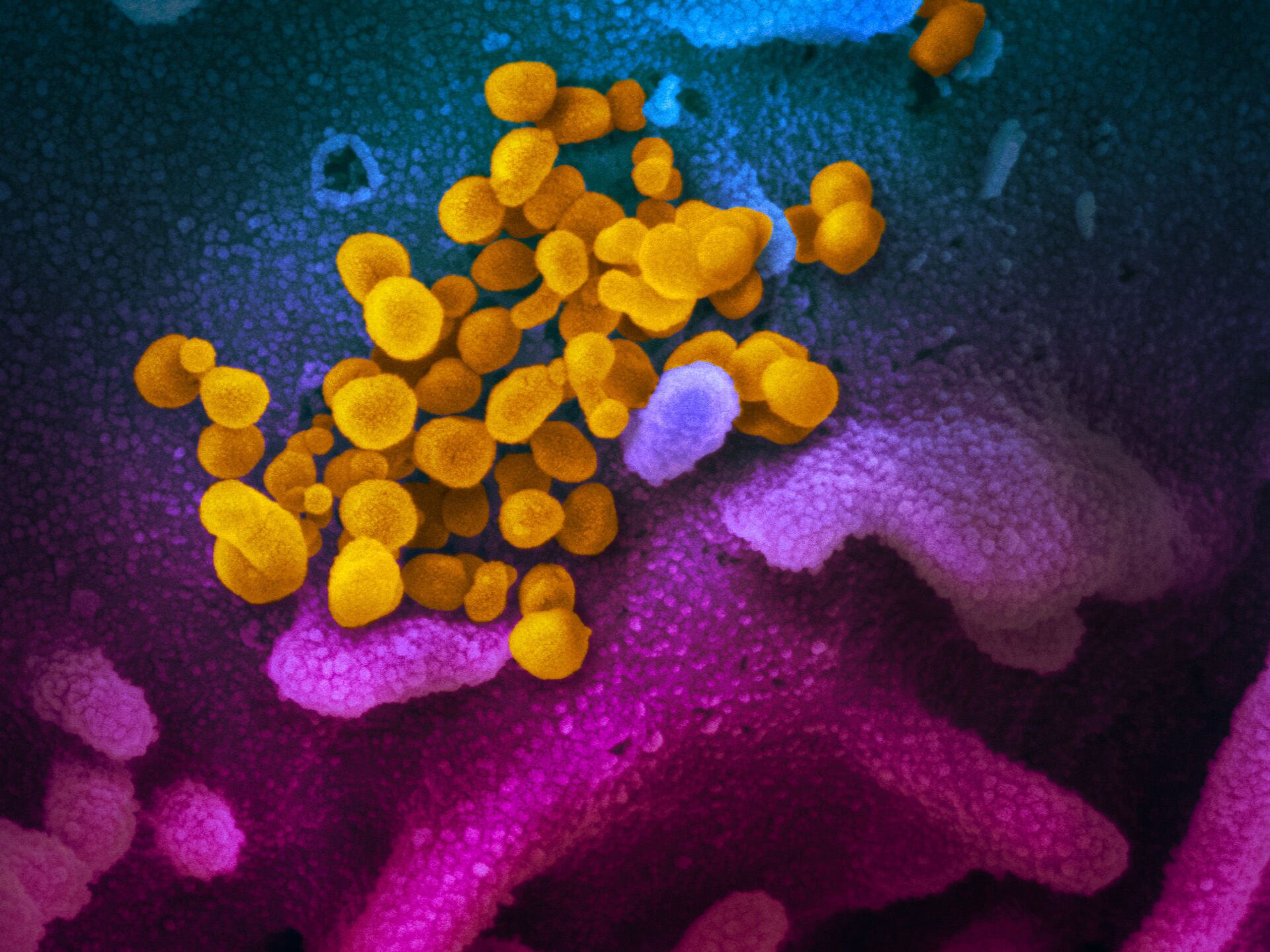
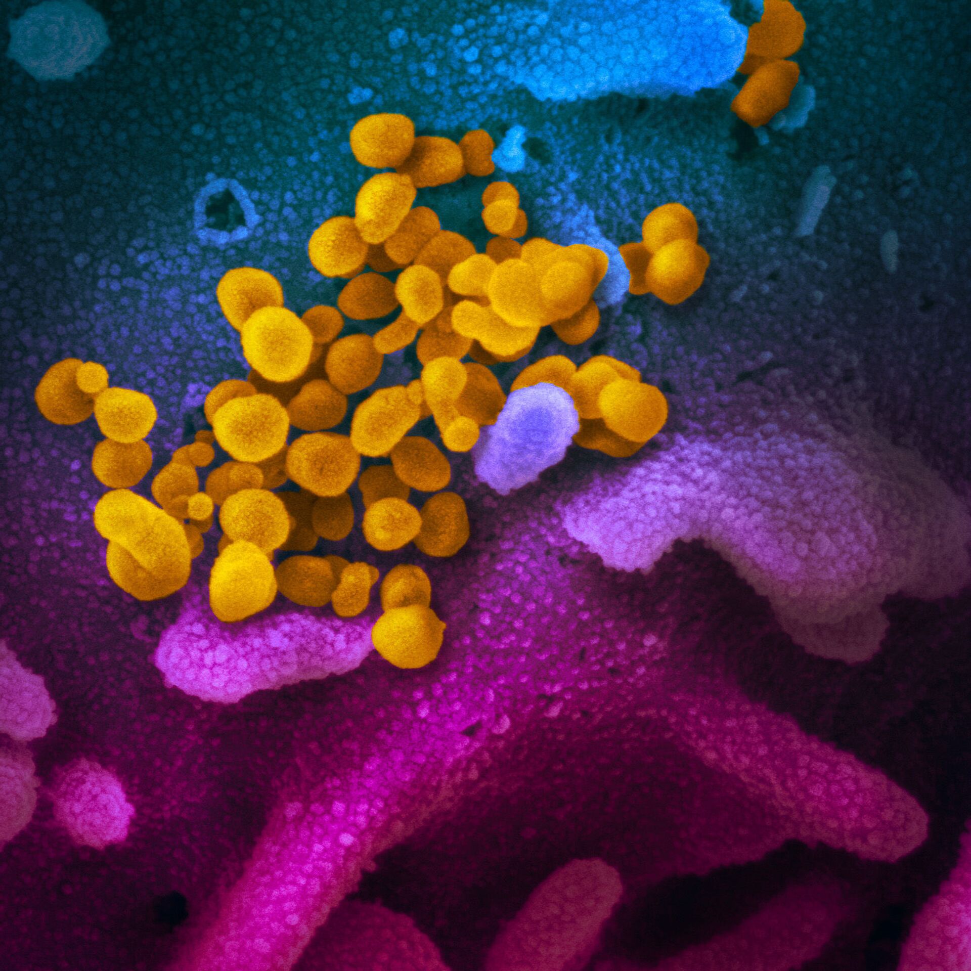
—also known as 2019-nCoV, the virus that causes COVID-19—isolated from a patient in the U.S., emerging from the surface of cells (blue/pink) cultured in the lab.")
Sputnik spoke to Dr Grace Roberts a virologist from Queen’s University Belfast, about different methods to study viruses.
Sputnik: Could you describe some of the different methods that scientists use to be able to examine viruses?
Dr Grace Roberts: I think what's really important when we talk about all these methods is they're all different and they've all got their advantages and disadvantages. But before I start explaining them all, I think it's important to keep in mind that they will use the different purposes essentially. So one of the common ones that we use a lot is what we call immunofluorescence and that's where we basically use antibodies and fluorescent attacks to kind of light up where viruses are going and ourselves.
This is quite a common technique that a lot of people use, because it's not too expensive. Most people have the microscopes available to be able to image stuff and it's quite quick, you can often do it within a day or two, so you can get results quite quickly from it. And that's really handy if you want to see basically where viruses are going in your cells and what proteins they might be interacting with within themselves. And then if you want more information you start having to go a bit more high resolution.
I talked a bit about super resolution microscopy, and that's very much, quite a new technique, that’s still being researched a lot so it's not as widespread and in use and it also needs a lot more high tech, microscopes that not everyone has access to. So that's more useful when you're talking about really pinpointing exactly where things are going in cells and what they might be interacting with, on a really, really small scale.
And then you might want to go past what is visually available and that's when techniques like electron microscopy and crystallography come in and they’re much more passively atomic scale. And so these are more useful if you're looking at really molecular interactions, you want to know exactly what molecules on the virus are interacting with what molecules on a cell and that can be useful in terms of things like drift design, so sometimes you want to design a drug that directly blocks some of these interactions.
So knowing the molecular detail of these interactions can mean that we can better design drugs to directly find and block and inhibit these kinds of things.
Sputnik: Regarding COVID-19, which method would be the most effective?
Dr Grace Roberts: So when we’re talking about testing in terms of getting people tested publicly, so, you know, do you have the virus or not? The tests that we use at the moment, the one that you take a swab for is a PCR test, so that's to detect the kind of genetics of the virus, and that is the most sensitive test to find out whether you've got a virus or not. And you know, that's pretty much your standard, and it is really sensitive, it can pick up tiny amounts of the virus. So it is worthwhile, but that doesn't visually show.
But you wouldn't normally use a visual technique just to know whether you've got a virus there or not in terms of public testing. We use a lot of techniques in the lab just to see, so sometimes we'll do an infection, say in the lab on some cells and we want to know that they're infected before we carry on. You can examine them on a microscope and this is done with a light microscope and you can see whether the cells look damaged or look sick.
It's normally quite obvious, especially with a virus, like COVID as it causes a lot of cell damage. So it's quite obvious when you look at healthy cells compared to infector cells, on a light microscope as even though you can't see the virus, that way, you can see the damage that it causes. So that's normally a really quick way that we can just check whether our cells are infected or not. And then it kind of depends what you want downstream from it. If you want to find out something really molecular, you might use a more advanced technique like electron microscopy that I talked about before, if you're just looking to see whether a virus is present, and in a certain cell type, maybe you could use immunofluorescence and you can find out really quickly and it will depend kind of what you want to know and using the best technique will tell you the information that you're looking for.

Dr Grace Roberts: Definitely electron microscopy and crystallography are far more advanced. So crystallography requires some really, really high tech expensive equipment, which is why I said earlier that, you know, we don't just have these kinds of things on our doorstep, you have to set up collaboration with people who have the materials and the machines to do this kind of stuff. And I couldn't even put a number on it to be honest with you how much that costs, but I know it's a lot.
Electron microscopes can cost hundreds of thousands to millions to buy. Things like fluorescent microscopes are much cheaper, you’re talking, ten to maybe hundreds or tens of thousands. But what's good about these things is, like I said, we often share with other institutions, you know, we have something that another institution doesn't, we will often let them use ours and then they'll let us use something of theirs that we don’t have. In order to like set up the samples of these techniques, that can be relatively cheap. So to produce the samples it normally doesn't cost that much money as you don't use that many resources, but the imaging can actually be quite expensive.


