
")
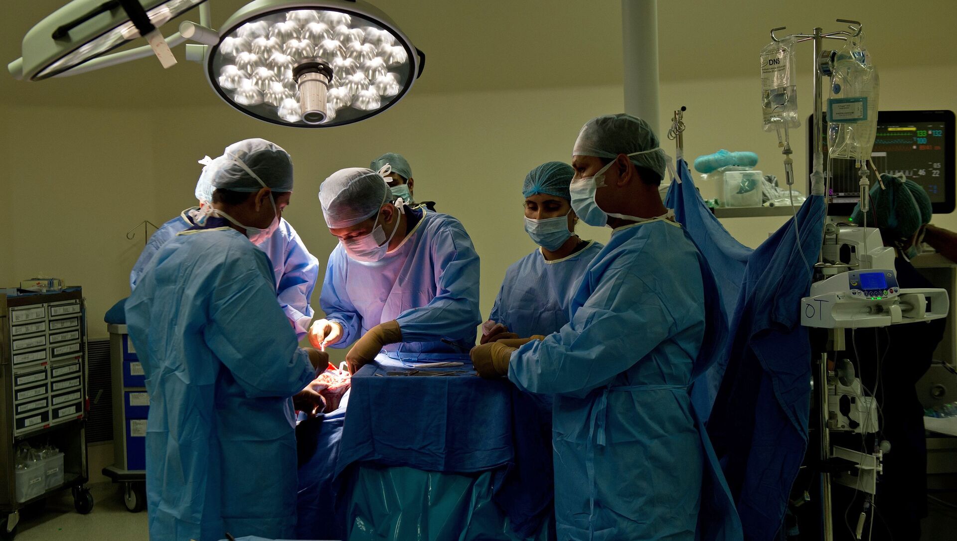
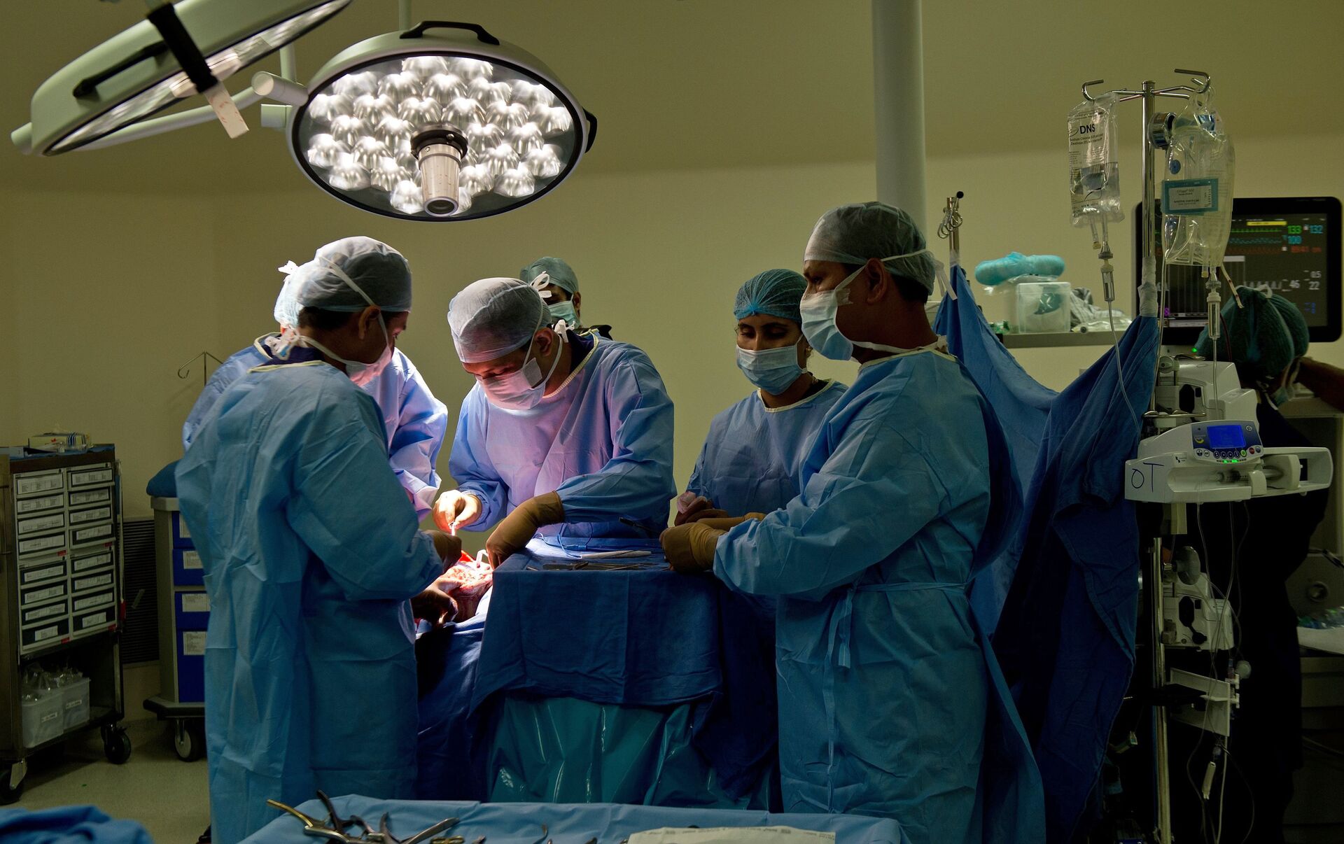
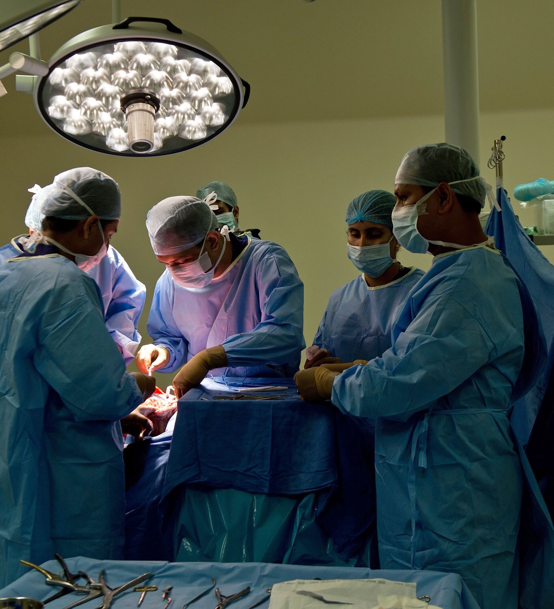
A team of doctors at a premier private medical college and research centre in India’s southern city of Kochi have carried out a rare surgical procedure on a 9-month-old child using 3D printing. The child was born with a very rare condition called complete cleft sternum.
“Narrow or small clefts can get away without surgery. But wide clefts, complete clefts with herniation of the heart and the great vessels are very dangerous and can cause sudden death even with mild trauma to the chest wall,” Dr Sandeep Vijayaraghavan, Professor of Plastic Surgery at the Amrita Institute of Medical Sciences and Research Centre in Kochi told Sputnik.
Dr Vijayaraghavan said, the team of doctors used a 3D CT scan of the region, simulated a mirror image using software and later used titanium to print the implant. This perfect titanium implant was sterilized and fixed in place in a simple procedure. “Previously we had to harvest ribs, split them and then screw them into place in a very complex and time-consuming procedure,” he added.
3D printing is a useful tool in surgical applications. It helps to understand complex deformities of the head and neck region, heart and chest wall, joints and bones. Various materials can be used for printing.
“In this 9-month-old child, we did a 3D CT scan and printed the ribs and the available cartilages that formed the edges of the cleft. We used the model and planned where the sternal edge should be cut to bring the edges together. We also planned the angle and how much of the cleft edges could be excised. We selected the best rib that could be used to bridge the sternal defect and the length, and the section of it was also decided on a model. We knew how much cartilage we could use to augment the rib graft,” said Dr Vijayaraghavan. “What we have done is not reported in any journal to date to the best of our knowledge.”
The team first did the procedure several times on a model to help decrease operating time and so that each team member knew what was expected of him.
Amrita Institute of Medical Sciences and Research Centre has been using 3D technology in complex facial bone reconstructions in both trauma and tumour surgeries. It is also the first hospital in India to have a 3D printing lab. The technique is especially useful in reconstructing the jaw bone with a bone from the leg. Dr Vijayaraghavan said they have also used this technique to reconstruct skull defects.


