
Scientists at the NIAID Integrated Research Facility (IRF) in Fort Detrick, Maryland have published photos of SARS-CoV-2, also known as the novel coronavirus, the virus that causes COVID-19.
To prevent the spread of the disease, multiple European countries have declared emergencies and introduced curfews, significantly slowing down economic activity, which is also resulting in lower levels of air pollution.
In the meantime, scientists across the globe are working on developing a vaccine against the coronavirus disease, which continues to kill thousands of people daily.

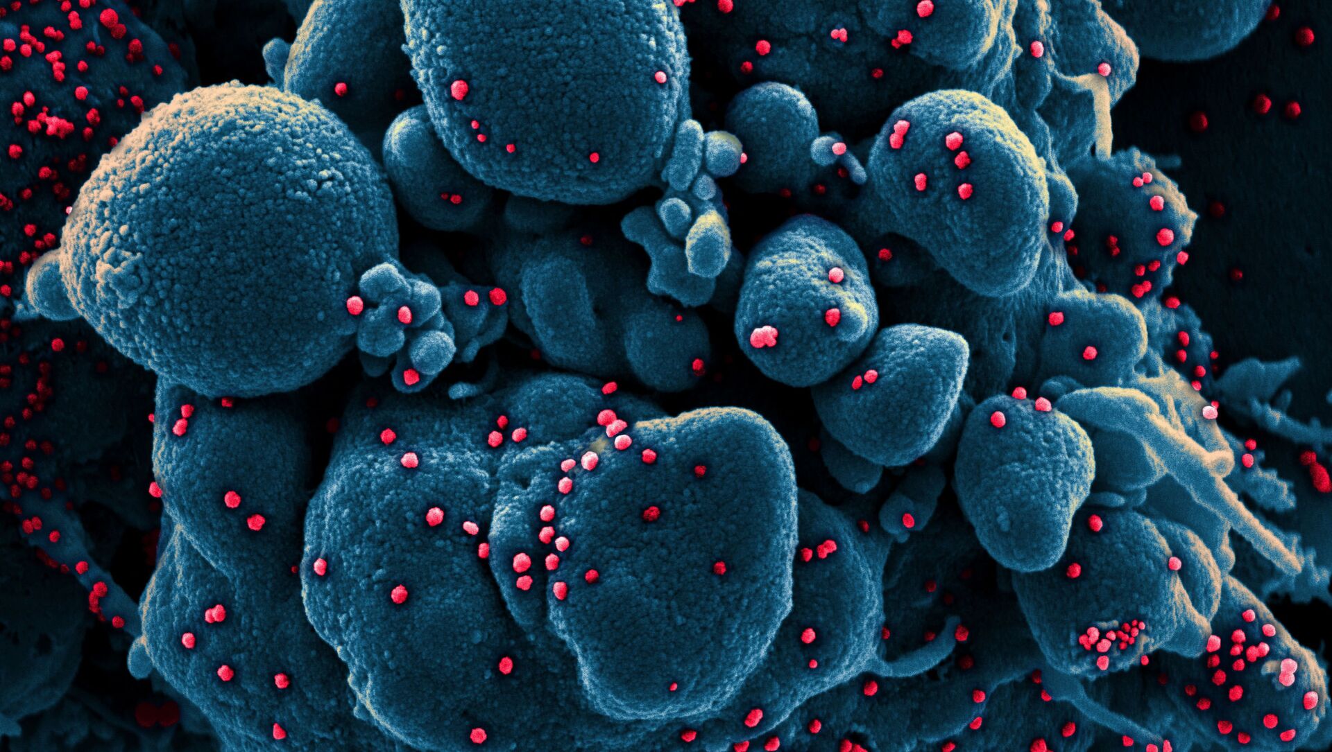
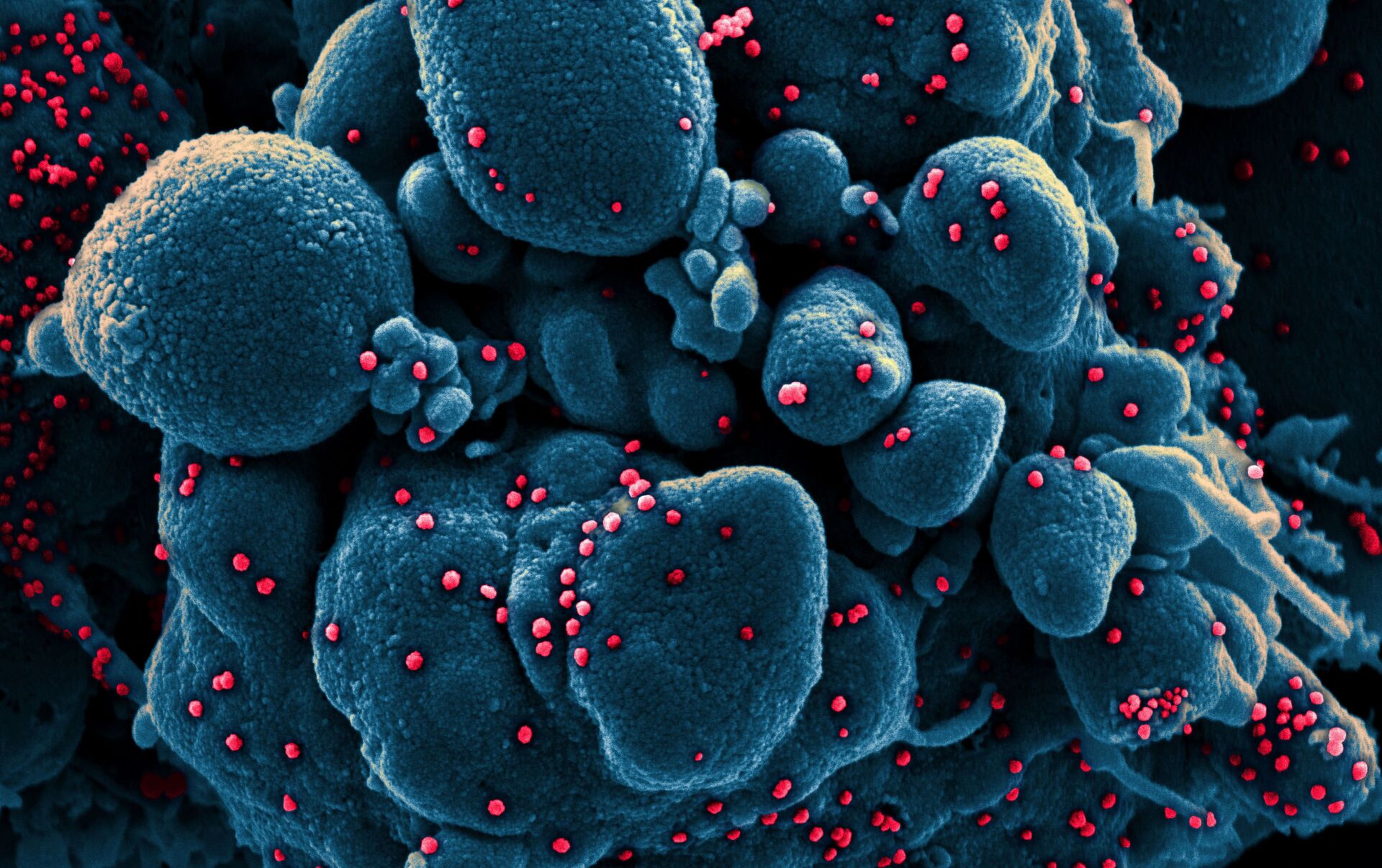
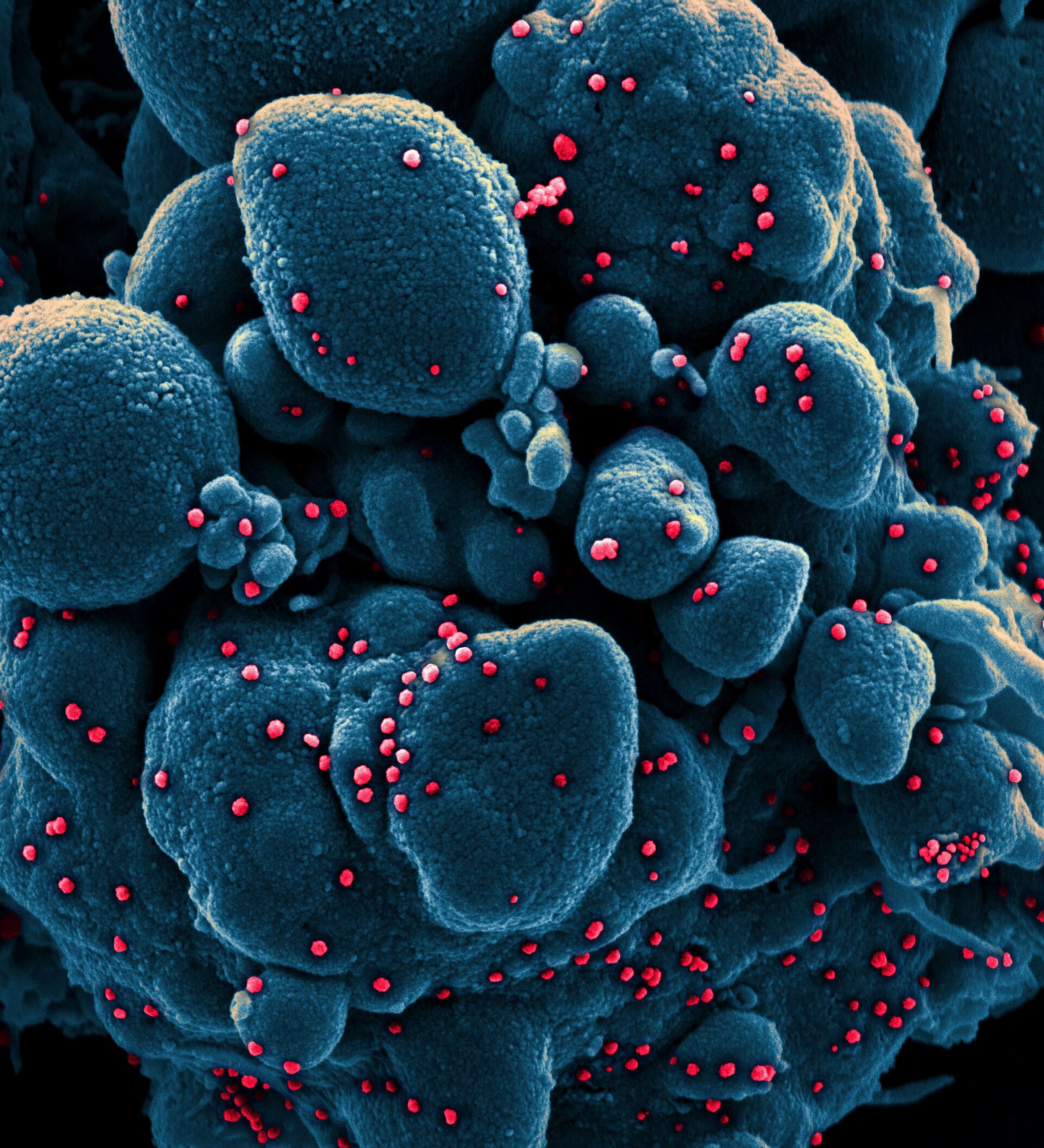
Colourised scanning electron micrograph of an apoptotic cell (blue) infected with SARS-COV-2 virus particles (red), also known as the novel coronavirus, isolated from a patient sample. Image captured at the NIAID Integrated Research Facility (IRF) in Fort Detrick, Maryland.

Colourised scanning electron micrograph of a VERO E6 cell (blue) heavily infected with SARS-COV-2 virus particles (orange), also known as the novel coronavirus, isolated from a patient sample. Image captured and color-enhanced at the NIAID Integrated Research Facility (IRF) in Fort Detrick, Maryland.

Colourised scanning electron micrograph of an apoptotic cell (blue) infected with SARS-COV-2 virus particles (red), also known as the novel coronavirus, isolated from a patient sample. Image captured at the NIAID Integrated Research Facility (IRF) in Fort Detrick, Maryland.

This scanning electron microscope image shows SARS-CoV-2 (round blue objects), also known as the novel coronavirus, the virus that causes COVID-19, emerging from the surface of cells cultured in a lab that were isolated from a patient in the US.

An undated transmission electron micrograph of the SARS-CoV-2 virus particles, also known as the novel coronavirus, the virus that causes COVID-19, isolated from a patient.

Colourised scanning electron micrograph of an apoptotic cell (greenish brown) heavily infected with SARS-COV-2 virus particles (pink), also known as the novel coronavirus, isolated from a patient sample. The image was captured and colour-enhanced at the NIAID Integrated Research Facility (IRF) in Fort Detrick, Maryland.

This scanning electron microscope image shows SARS-CoV-2 (yellow) – also known as 2019-nCoV, the virus that causes COVID-19 – isolated from a patient in the US emerging from surface of cells (blue/pink) cultured in a lab.

An undated transmission electron micrograph of SARS-CoV-2 virus particles, also known as the novel coronavirus, the virus which causes COVID-19, isolated from a patient.

An undated colourised scanning electron micrograph of an apoptotic cell (green) heavily infected with SARS-COV-2 virus particles (purple), also known as the novel coronavirus, the virus which causes COVID-19, isolated from a patient sample.


