
This method allows for the rapid assessment of the toxicity of chemotherapeutic agents used for treating cancer, which results in a more accurate survival prognoses. The development of this new nanoelectrode is also important for the pre-clinical trials of various drugs, as it allows performing necessary tests on single living cells rather than laboratory animals. The results of the new research were published in the Scientific Reports journal.
READ MORE: Sorry ET, Got Here First: Russian Scientist Suggests Humans Would Destroy Aliens
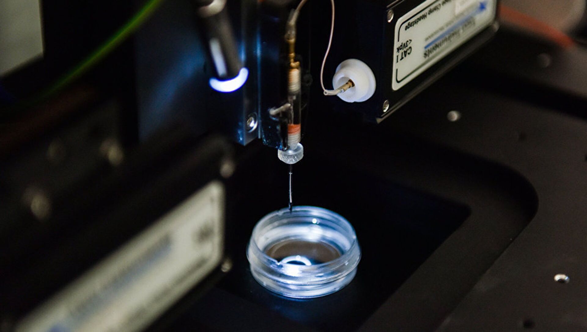
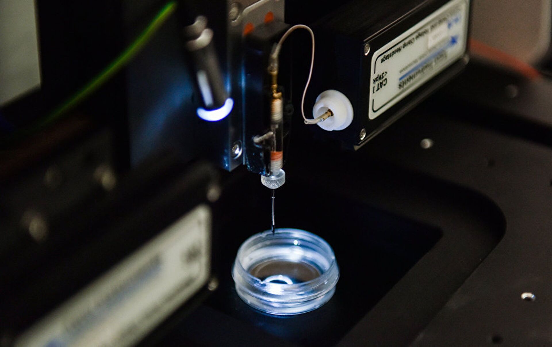
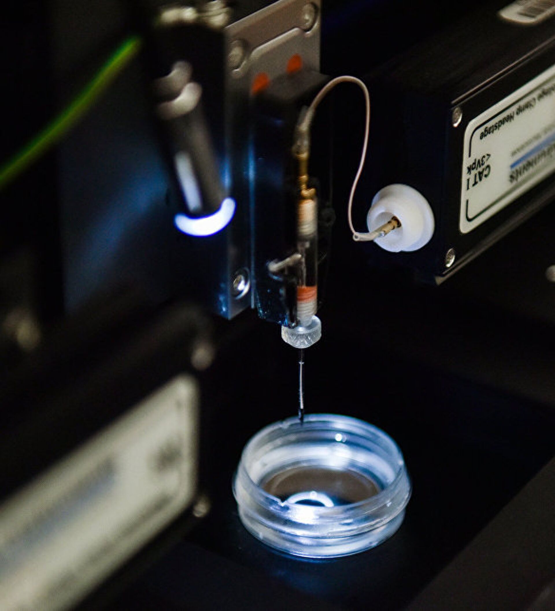
Until now, there was no method for assessing the level of intracellular toxicity without destroying the cell. With the participation of the National University of Science and Technology MISiS (NUST MISiS), the international team of researchers decided to develop such a method by using nanocapillaries, which are traditionally used in scanning ion-conductance microscopy. To achieve this goal, they upgraded a nanocapillary by filling it with carbon and platinizing the tip, thus creating a nanoelectrode that can be used to probe the cell. It can also be introduced inside the cell through a nanopuncture, which heals in an instant as soon as the nanoelectrode is removed.

Magnetic nanoparticles are widely used for targeted drug delivery and as contrast agents in magnetic resonance imaging, induced hyperthermia (for “burning out” tumors from the inside) and magnetic field assisted radionuclide therapy.
Researchers need to be sure that the increasing application of these particles does not damage a patient’s health, thus it is important to develop methods that can ensure the high efficiency of bioreaction analysis and the accuracy of the toxicity prognosis.
READ MORE: Russian Scientists Develop Express Test to Identify Cancer
It is known that magnetic nanoparticles cause the generation of reactive oxygen species (ROS) inside cells. ROS are oxygen ions, free radicals and peroxides formed as a result of natural metabolism or under the effect of ionizing radiation. The high cellular levels of ROS usually result in oxidative stress and damage to the cells due to oxidation. This means that the increased content of ROS is one of the first signs indicating the presence of intracellular toxicity. NUST MISiS researchers used this effect to assess the toxicity of magnetic nanoparticles based on measuring ROS content.
“We have developed a stable probe for measuring intracellular ROS using a platinized carbon nanoelectrode,” said Alexander Yerofeyev, one of the authors of the research and senior researcher at NUST MISiS Biomedical Nanomaterials laboratory, in his interview with RIA Novosti. “The results of measuring ROS levels obtained using this new method differ significantly from the results obtained with traditional methods. Outdated technology fails to detect the corresponding difference in ROS levels.”

Scientists also emphasize the importance of and prospects for using nanoelectrodes for this type of analysis. They believe that this will allow performing a relatively fast, sensitive and efficient assessment of the toxicity level of nanoparticles in single cells.


