
The trials are being led by Jason G. Newman, an associate professor of Otorhinolaryngology — ear, nose and throat medicine — at the University of Pennsylvania. This is the first time surgeons have used intraoperative molecular imaging, which utilizes an injectable dye engineered to accumulate in cancerous tissues in patients with neck and head cancer, ScienceDaily.com reported.

Newman and his team are using a specific contrast agent, known as "indocyanine green," a fluorescent dye approved by the US Food and Drug Administration years ago for medical diagnostics.
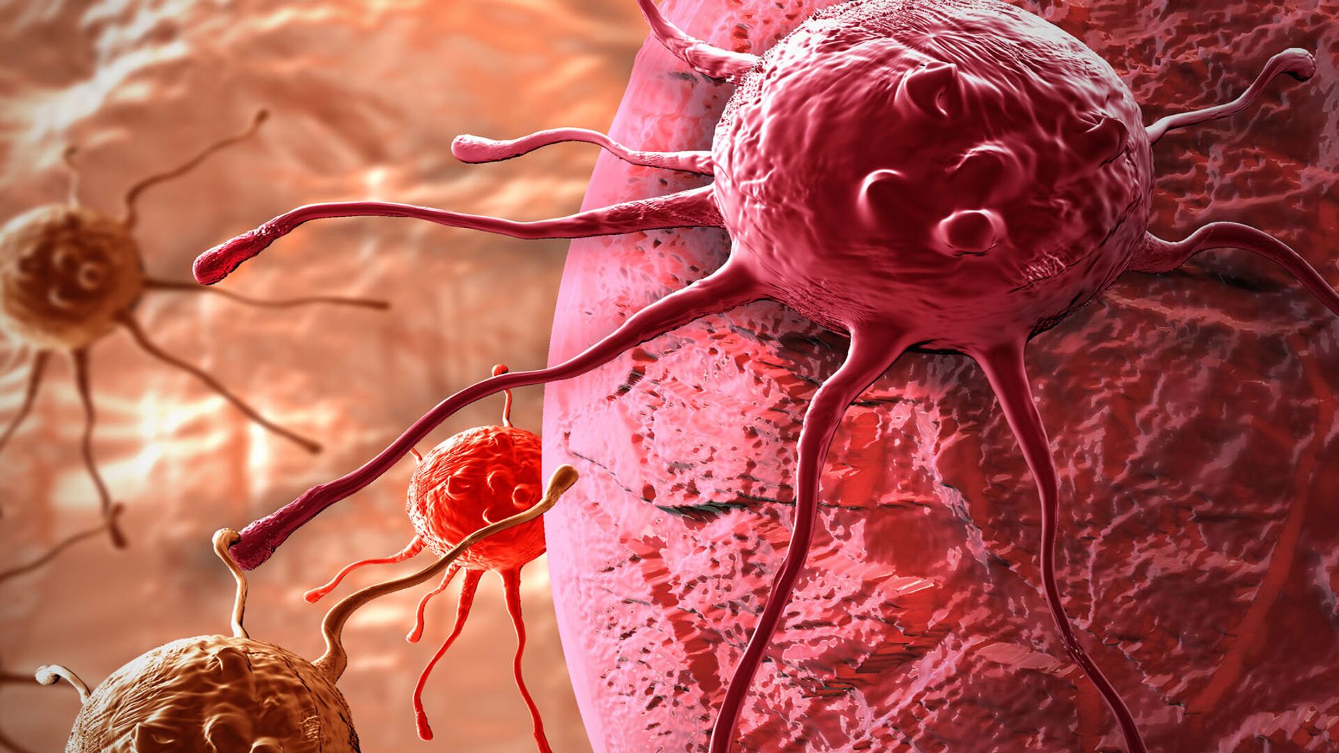
When patients are injected with the dye, cancer cells that may not be visible in CT scans "glow" under near-infrared light because the dye has been specially engineered to be attracted to the tumor. The dye only begins to glow after it is activated when exposed to light during surgery, according to the Penn Medicine website.
In an interview with Healio, Newman explained that the trial will consist of patients coming into an infusion suite a day before their surgeries, where they will be injected with indocyanine green in order to determine which tumors of the head and neck respond best to this imaging technology.
"We have patients with different tumors of the head and neck and we are in the early stage of determining which tumors are the most appropriate on which to use this imaging technology. We do not know where the ‘sweet spot' of this technology is in the head and neck arena," Newman explained in the interview last month.
"They [the patients] get injected, go home and come back the next day for surgery. The only difference is that we use a camera to image the tissue, and we determine the utility of the fluorescent dye. We are not yet using this to make formal decisions during surgery, but we are using intraoperative molecular imaging to help determine what role the market should or can play in the management of these cancers," he added.
When asked about the expected time for results, Newman noted that they have already had abstracts accepted for publication in scientific journals. However, the timeline for the trials is "ongoing."
"The timeline is ongoing because — unlike a drug trial, where we'd have to wait for efficacy — we know the efficacy at the time of surgery and pathology. The most interesting information comes in that ‘sweet spot' when tissue looks normal to the naked eye, but then imaging shows the tissue is fluorescing, we test it and we start to determine that the technology is doing a better job than our eyes distinguishing tumor from normal tissue," Newman noted.

Although surgery has long been used to successfully cure cancer, stray tumor cells can be left undetected. In addition, while surgeons often remove potentially suspicious tissue during surgery, this does not come without the risk of also damaging healthy tissue in the process.
According to the National Cancer Institute, head and neck cancer make up nearly 4 percent of all cancers in the US. Around 75 percent of these types of cancer cases are caused by smoking and drinking, although an increasing number of cases are being connected to human papillomavirus (HPV), an infection that causes warts in various parts of the body.
Newman and and his team are not the first to delve into intraoperative molecular imaging for cancer detection. Sunil Singhal, a doctor at the Abramson Cancer Center's Center for Precision Surgery at the University of Pennsylvania, was also recently inspired to develop glowing tumor dye technology, which he dubbed TumorGlow. However, Singhal has only tested the technology in patients with lung and brain cancer.
"[With dyes] it's almost like we have bionic vision," said Singhal. "We can be sure we're not taking too much or too little." His hope is that TumorGlow and other similar technologies will become a standard of care when surgically resecting cancers.


