New DNA Imaging Technique Shows Chromosomes in High-Res

© REUTERS / National Human Genome Research InstituteA DNA double helix is seen in an undated artist's illustration released by the National Human Genome Research Institute to Reuters on May 15, 2012
A DNA double helix is seen in an undated artist's illustration released by the National Human Genome Research Institute to Reuters on May 15, 2012 - Sputnik International
Subscribe
Researchers have come up with a new method of imaging DNA, which enables scientists to better examine damage or changes to the molecules.

Neanderthal man - Sputnik International
New DNA Sequencing Reveals Prehistoric Man's Dramatic Journey Round Europe
Researchers have developed a new DNA imaging technique that is able to examine the structure of individual DNA strands, which measure just a few nanometers across, in much more detail than the methods currently available. 

This new method could allow scientists to gain important insights into what goes wrong when DNA becomes damaged, The Optical Society reported on Friday.

The research, published in the peer-review journal Optica, was led by W. E. Moerner of Stanford University, who carried out the first optical detection and spectroscopy of a single molecule in 1989. 

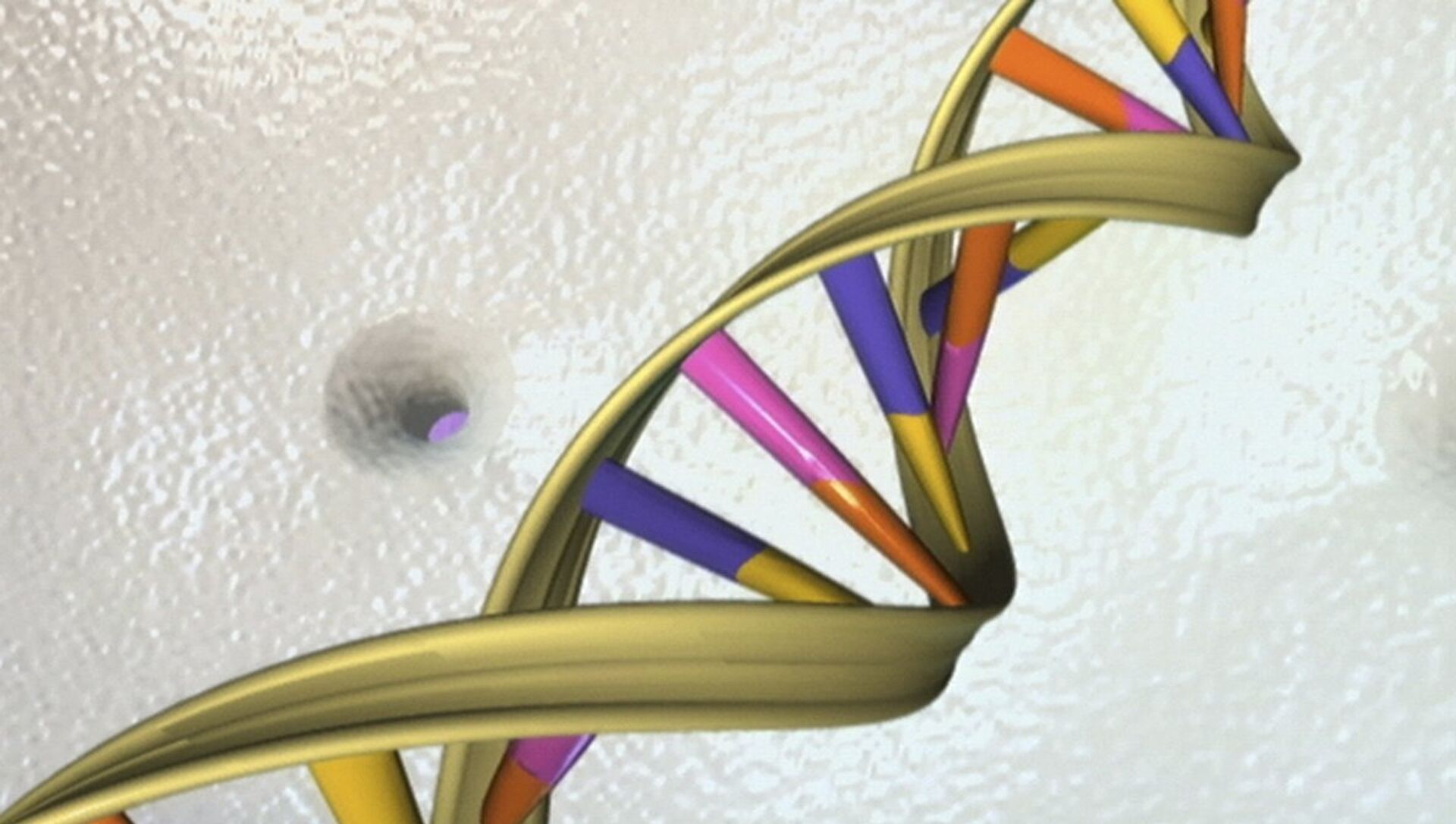
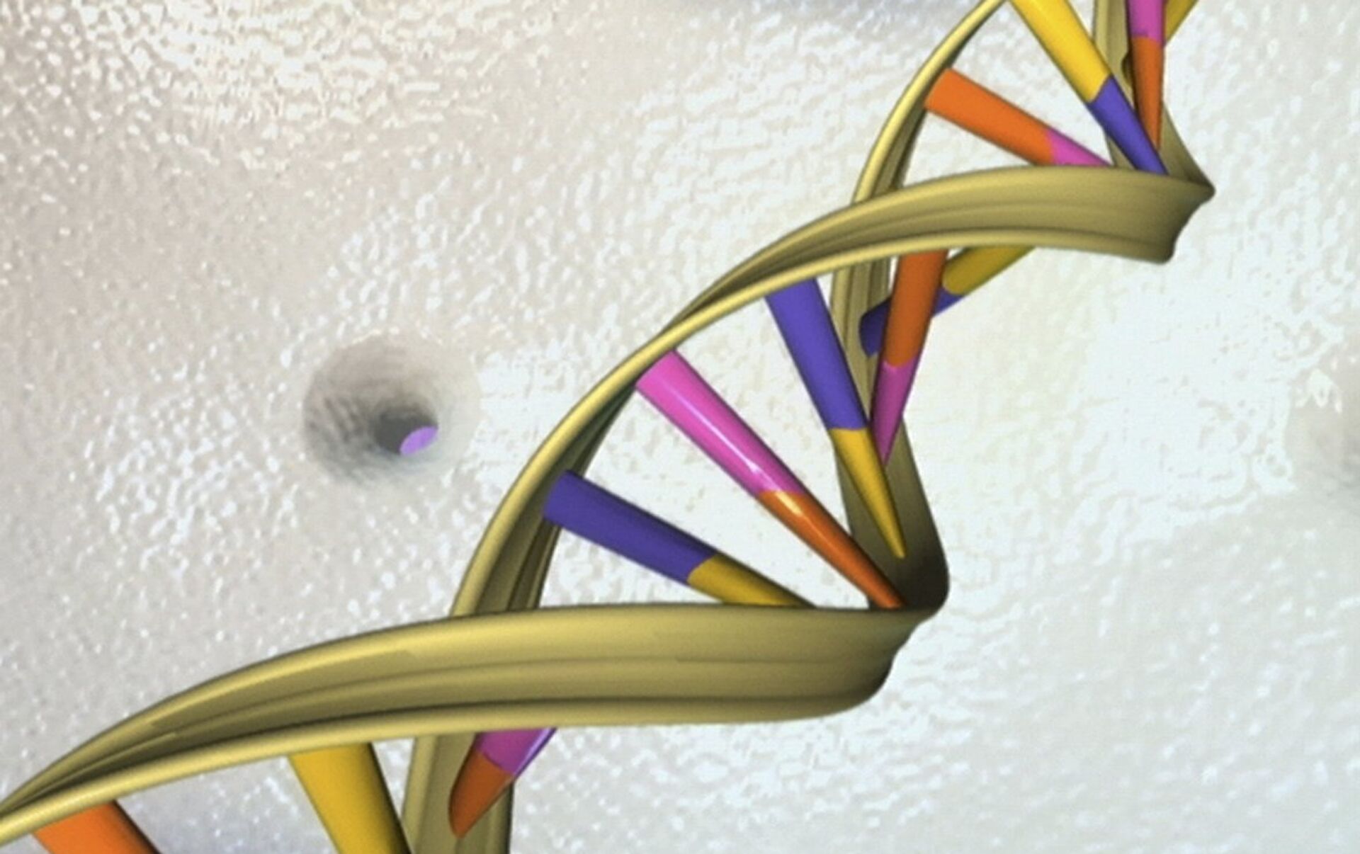
The team has improved this technique by adapting the laser used to analyze thousands of fluorescent dye molecules, which can slide into the areas between the DNA base pairs, attach to the side of the DNA, or connect to it via a floppy tether.

By augmenting this conventional fluorescence microscope with an electro-optic modulator (EOM), they were able to observe the orientation and movement of the dye molecules.

© osapublishing.orgSuper-resolved images and single-molecule orientation measurements acquired using the intercalating dye SYTOX Orange
Super-resolved images and single-molecule orientation measurements acquired using the intercalating dye SYTOX Orange - Sputnik International
Super-resolved images and single-molecule orientation measurements acquired using the intercalating dye SYTOX Orange
In a typical imaging experiment, they acquire up to 300,000 single molecule locations and 30,000 single-molecule orientation measurements in just over 13 minutes.

"You can think of these new measurements as providing little double-headed arrows that show the orientation of the molecules attached along the DNA strand," Moerner said

"This orientation information reports on the local structure of the DNA bases because they constrain the molecule. If we didn't have this orientation information, the image would just be a spot."

HIV-infected H9 T cell - Sputnik International
World
Cure Around the Corner: In First, HIV Virus DNA Removed From Living Tissue
The scientists say that nanoscale information provided by the new technique could be useful for monitoring changes in DNA or damage to particular regions of the molecule. It could also be used to monitor interactions between DNA and proteins, which drive many cellular processes.

"If someone has a single-molecule microscope, they can perform our technique pretty easily by adding the electro-optic modulator," said Adam Backer, lead author of the paper.

"We've used fairly standard tools in a slightly different way and analyzed the data in a new way to gain additional biological and physical insight."

Newsfeed
0
To participate in the discussion
log in or register
loader
Chats
Заголовок открываемого материала